Product Details
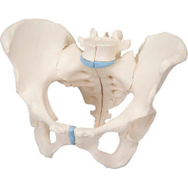
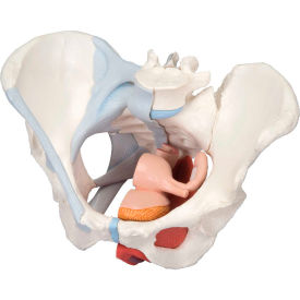
This life-size four-part model of a female pelvis provides an in-depth representation of the topography of bones, ligaments, pelvic floor muscles, and female pelvic organs. The right half illustrates the bones along with pelvic ligaments, while the left half exhibits the pelvic floor muscles, including levator ani, ischiocavernosus, deep and superficial transverse perineal, external anal sphincter, and external urethral sphincter. It features a partially removable bulbospongiosus that highlights the vestibular bulb and Bartholin gland. Additionally, a removable midsagittal section through the urinary bladder, vagina, uterus, and rectum reveals the anatomical relationships to the pelvic floor muscles at the openings for the urethra, vagina, and rectum. This model is ideal for gynecological studies and detailed anatomical exploration.